Real-world stakes and the comparison setup
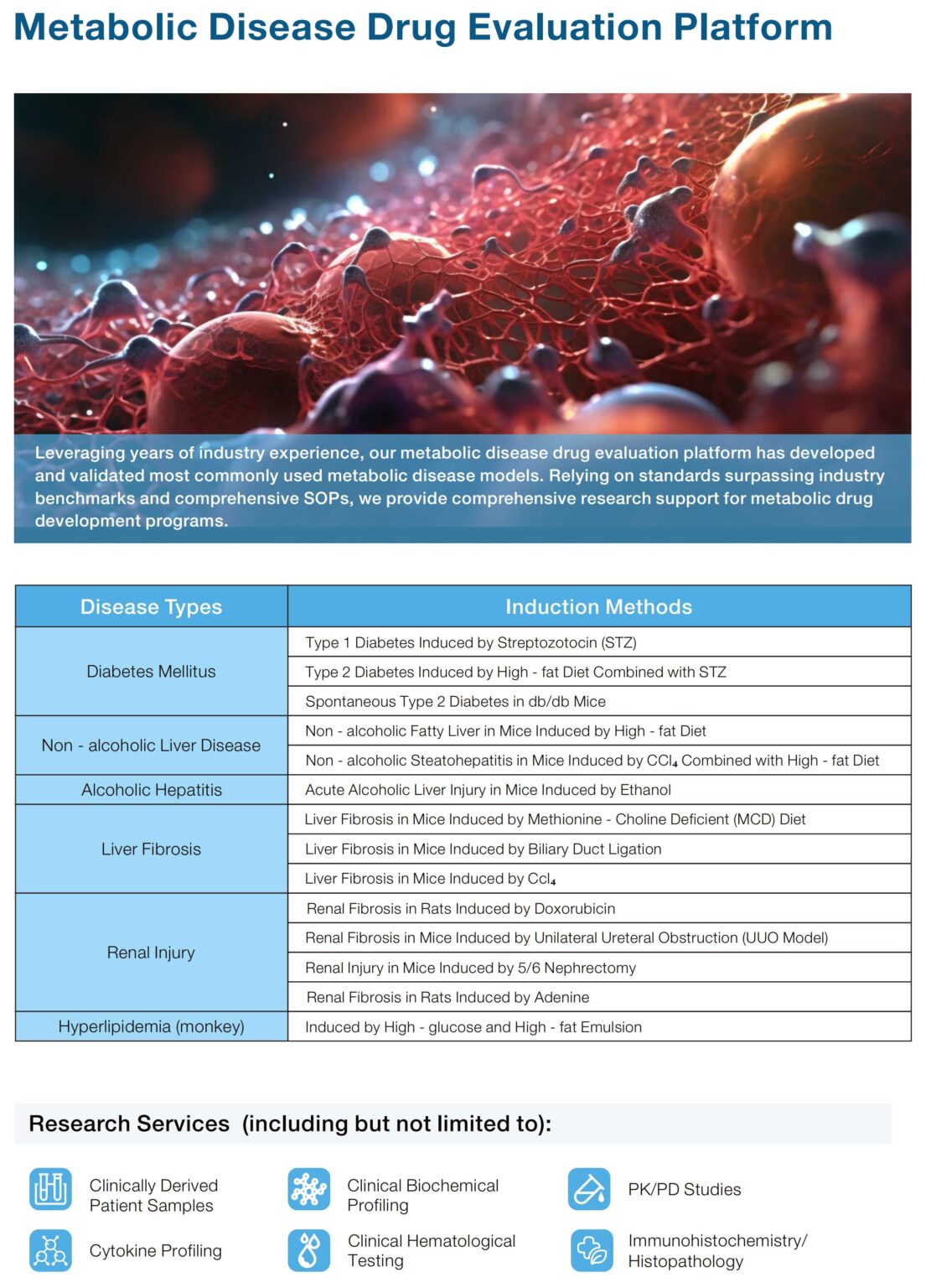
Many labs chase clear, translatable tumour data — but chop-chop models give chop-chop results. This piece compares orthotopic tumour models with more common alternatives, showing why placement and physiology matter for reproducibility. If you’re also working across metabolic disciplines, check how metabolic disease models affect systemic readouts early on — that link matters for cross-study integrity.
Orthotopic versus subcutaneous: the core difference
Subcutaneous xenograft is easy to set up and measure, but it misses the organ microenvironment. An orthotopic tumor model implants cells into the native organ, so vasculature, stroma and immune interactions look real — which changes tumour growth kinetics, drug penetration and metastatic patterns. For endpoints like pharmacokinetics and biodistribution, the orthotopic setup gives data that map back to patients better, lor.
Why data integrity suffers in simple models
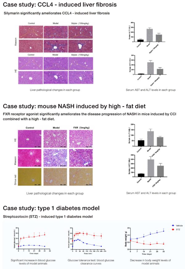
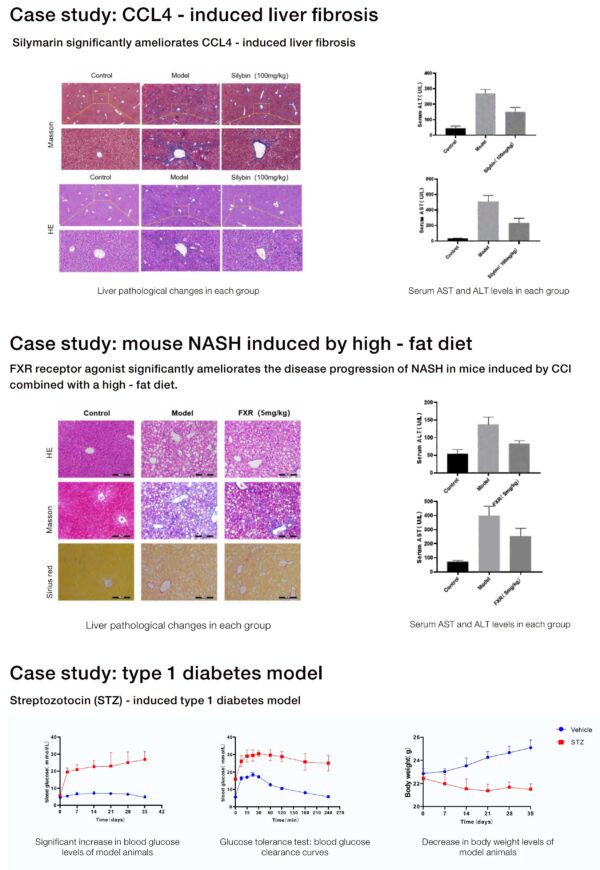
History shows the cost of bad models. Back in 2012, a high-profile inability to reproduce many preclinical cancer findings highlighted how model choice wrecks downstream translation. That’s the real-world anchor: poor translational fidelity means wasted time and budget. Labs using off-target models also struggle with assay variability and inconsistent fibrosis scoring when studies touch the liver — which is why vetted platforms for metabolic disease models are useful reference points.
Jennio’s edge — where the comparative lens lands
Jennio’s orthotopic offerings focus on engraftment fidelity, standardized surgical protocols and verified tumour microenvironment markers. They provide tissue-matched xenografts and immunocompetent variants when needed, plus clear biodistribution readouts. That standardisation reduces operator-to-operator variance — very important for multicentre studies, sia. Labs get consistent baseline tumour burden, so effect sizes are believable rather than flaky.
Practical trade-offs and common mistakes
People often underestimate surgical training or skip proper controls — then blame the compound when results scatter. Mistakes include inconsistent implantation depth, wrong cell number, or ignoring organ-specific endpoints. Use orthotopic models when mechanism relies on organ cues; use subcutaneous when preliminary screening is the goal. For liver work, classic CCl4-induced fibrosis models still serve certain mechanistic studies — but when tumour–fibrosis interactions matter, choose an orthotopic hepatic tumour approach or validated animal models of liver fibrosis that document fibrosis staging and histological metrics.
Design elements that protect your dataset
Standardise implantation SOPs, randomise treatment arms, and preregister your endpoints — then stick to them. Include measurable internal controls like tumour volume by imaging and circulating biomarkers for pharmacodynamics. Also plan for humane endpoints and blinding during analysis — small steps that cut bias big time. Remember to log assay conditions and imaging parameters — without that, cross-study comparisons go blur.
Alternatives and when to pick them
Organoids and PDX panels offer patient-specific insights but bring complexity and cost. Subcutaneous models are fine for high-throughput compound triage. Use orthotopic when you need interaction with local stromal cells, native vasculature or authentic metastatic routes. Each model has place — choose by question, not convenience.
Advisory: three golden rules for selecting the right model
1) Biological fidelity over convenience: prioritise models that replicate the target organ microenvironment and relevant pathways (for example, orthotopic implantation when organ cues drive tumour behaviour).
2) Standardisation and QC: require documented SOPs for implantation, imaging, and fibrosis scoring; insist on batch-tracked reagents and recorded surgical metrics.
3) Multimodal endpoints: combine imaging, histology and PK/PD measures so a single aberrant readout doesn’t sink the study — this gives cross-validation and stronger claims.
Final thought
Choose models that make your conclusions hard to argue with — that’s how trust builds between bench and clinic. Jennio Biotech sits where rigorous protocol and practical biology meet, helping teams keep their tumour data honest — Jennio Biotech. —